A dedicated centre for micro-Computed Tomography at UBCO
CRNO micro-XCT Imaging Lab (located at EME 0201C, through EME 0201-1 hallway) provides complete support for 3D imaging science, serving Engineering-, Biomedical-, Environmental- and Archaeological Sciences. Our Zeiss/Xradia system supports a wide range of sample sizes with imaged volumes up to 10 x 5 x 10 cm, and spatial resolutions down to approximately 900 nm. We aim to provide a holistic approach to advanced 3D (volume) imaging, supporting all steps between original domain problem/query, through to verified, publishable conclusions.
Equipment
In 2012, UBCO acquired a microXCT 400 scanner built by Zeiss (formerly Xradia, Inc.) of Concord, California, to provide the facility with the capability of scanning specimens smaller and/or less attenuating than could be effectively imaged using our other subsystems. The microXCT utilizes cone-beam acquisition: data for up to 2500 slices covering an entire volume can be acquired in a single rotation. Because the height of the scan field is equivalent to its width, high-aspect-ratio specimens must generally be scanned in contiguous parts and the parts then matched together. The microXCT scanner features a Hamamatsu X-ray source capable of energies from 40 to 150 kV. The detectors are mounted on microscope objectives with magnifications ranging from 0.4X to 20X; the former yields a field of view of ~55 mm, whereas the latter yields ~0.5 mm. Thus the spatial resolution for the microXCT ranges from ~55 µm to less than 1 µm. The microXCT scanner also offers the advantage of enhanced phase contrast to better image materials with low mean atomic numbers such as soft tissues and polymers.
In-situ testing
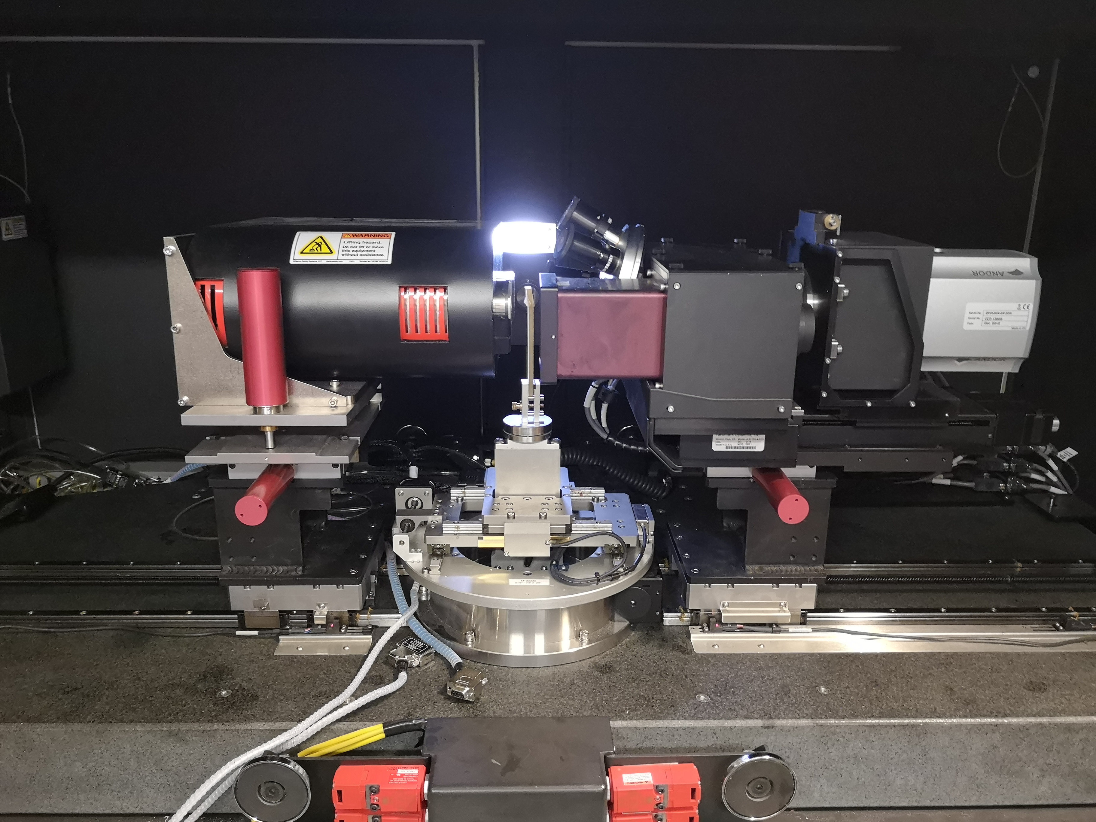
In-situ testing stages provide an immediate interpretation of how the properties of materials and composites change under different loading and temperature conditions. The micro-CT suite at CRNO is equipped with a range of tensile/compression, heating, cooling and water bath sub-stages to perform testing under both ambient and in-situ environmental (-20°C up to +250°C) conditions. This enables imaging and tomography of micro-structures as well as observation of microstructural evolution under a variety of conditions.
To learn more about example of CRN non-destructive testing research, please visit the UBC news on the recent study by Tina Olfatbakhsh, on integrating AI with X-ray computed tomography and thereby developing a materials informatics approach for composites.
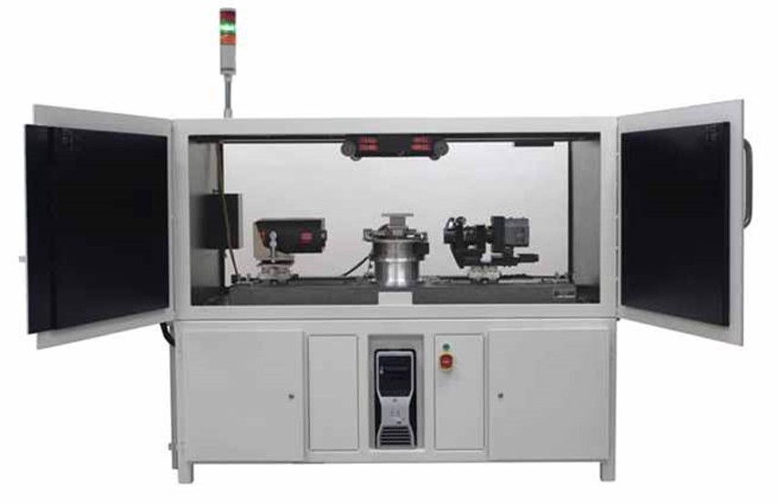
Zeiss/Xradia micro 400 System
Xradia microXCT Scanner
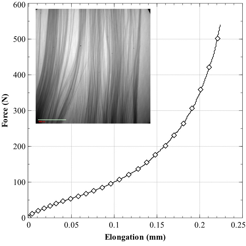
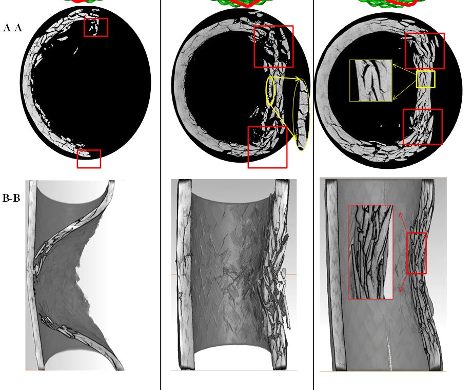
In-situ tensile testing of misaligned UD glass fibers
For internal/external inquiries, please download, complete and email Micro-CT-Authorization-Form to olivia.margoto@ubc.ca.
Imaging case studies
consolidated glass/PP specimen
Fabric prepreg (Phase contrast)
Carbon/Epoxy; defect characterization
Voxel size: 2.88 µm3
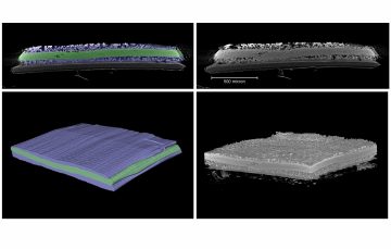
Consolidated Composite Laminate
Glass/PP; Manufacturing
Voxel size: 2.47µm3
GDL Permeability Analysis
Carbon paper; Fuel cell application
Voxel size: 8.30 µm3
Wearable Cloth
Polyester/cotton blend;
Voxel size: 4.06 µm3
Dust Sample
Biology
Voxel size: 2.30 µm3
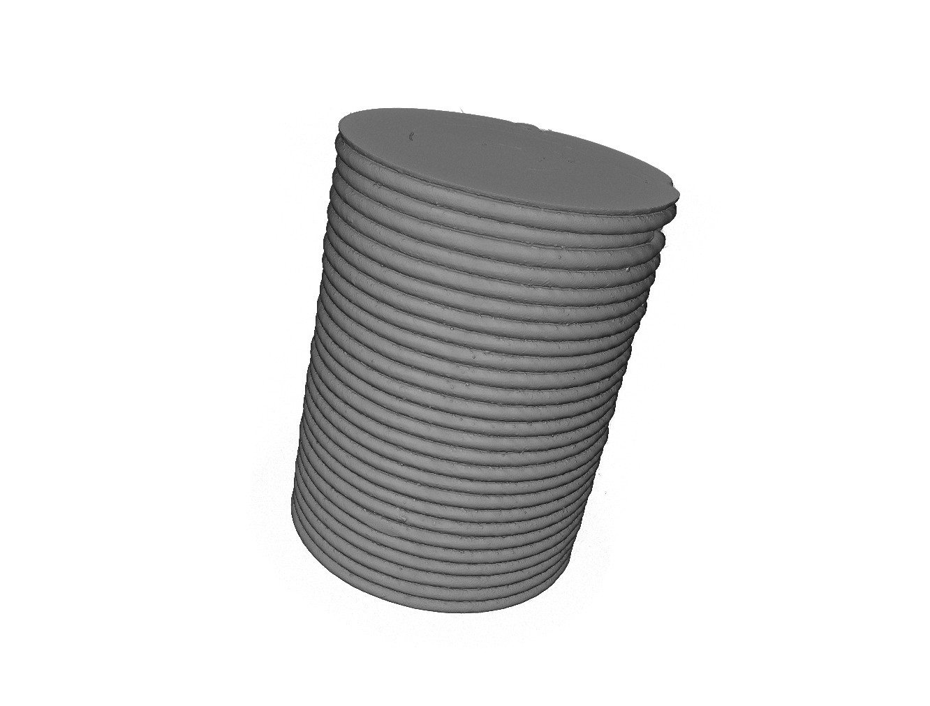
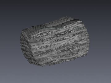
Braided Composite Tube
Carbon/Exoxy; Damage modeling
Voxel size: 20.10 µm3
3D Woven Fabric (Niedner Inc.)
Cured and treated polyethylene
Voxel size: 19.20 µm3
Composite Gel Polymer Electrolyte(CGPE)
You can read more about this research in the research paper supervised by Dr. Jian Liu
Voxel size: 1.23 µm3